

A girl born in 2002, is ill since May 2015 with headache, weakness, drowsiness. In September 2015 addressed to local neurosurgeon. At MRI of brain a tumor sized 25x20x20 mm is seen in the projection of the hypothalamus, with possible 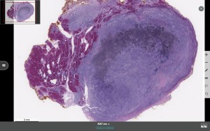 penetration into the area of the 3rd ventricle’s bottom. It has heterogeneous structure and rather sharp but irregular contours. For further examination the patient directed to the endocrinologist. At the thyroid ultrasound investigation, a hypoechoic inhomogeneous solid structure sized 23x43x24 mm which occupies the entire thyroid’s right lobe with transition into isthmus is found. Structurally similar lesions are found in the left lobe of the thyroid gland, with the size of 14×13, 16×8 and 6×6 mm.
In the proximity to the lower pole of the thyroid gland multiple lymph nodes are visualized, with the largest size of 13×6 mm. In the early October 2015 thyroidectomy with lymph node dissection on both sides was performed.
To see the proper diagnosis please register here: